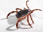
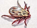
Electron microscope images
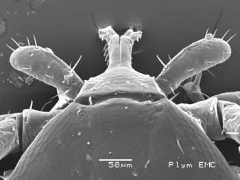
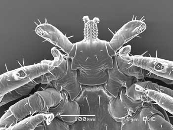
Sheep tick - Ixodes ricinus
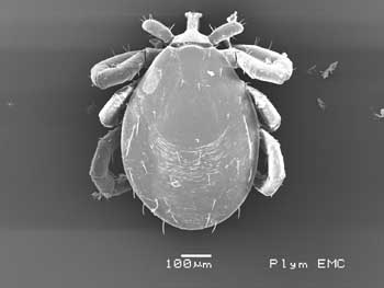
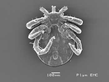
LARVA - note 6 legs
Click here for:
larva, nymph,
male, female,
mating ticks and
comparing mouthparts photographs

These photos were taken on the JEOL 5600LVSEM
scanning electron microscope at the University of Plymouth Electron Microscopy
Centre on 10th August 2009.
 |
 |
|
Dorsal view of a larva |
Ventral view of a larva |
 |
 |
|
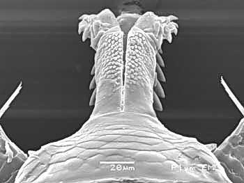
Dorsal view of the capitulum |
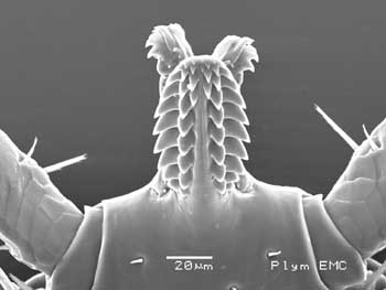
Ventral view of the capitulum |
 |
 |
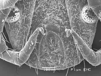
Dorsal view of the hypostomal region,
showing
the cheliceral sheathes and chelicerae |
Ventral view of the hypostome, showing
the
chelicerae extended dorsally beyond |
 |
 |
|
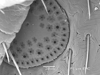
Spiracles and anus |
Spiracle, detail (backscattered electron
image) |
 |
 |
|
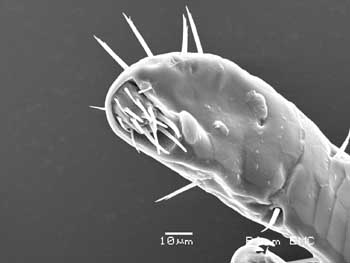
Pedipalp tip, showing terminal sensory
organ |
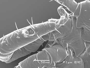
Right leg 1, showing Haller's organ |
 |
 |
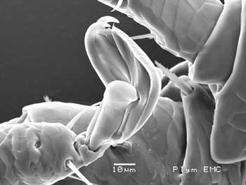
Left foot 1, note the branched seta at
right, almost
touching the foot, possibly a sensory structure? |
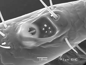
Haller's organ, detail |
Acknowledgement: My thanks to the
staff of the University of Plymouth Electron Microscopy Centre, Dr Roy Moate,
Peter Bond and Glenn Harper, for enabling me to take these photographs.