 |
 |
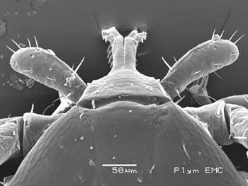
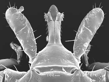
Larva - dorsal view of the capitulum,
chelicerae extended |
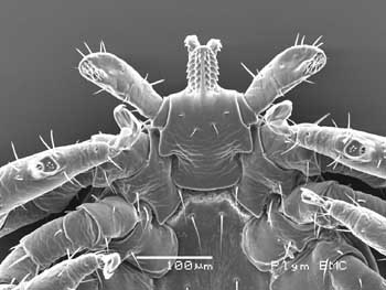
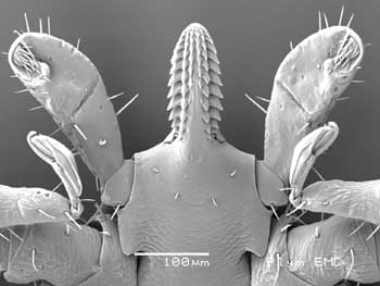
Larva - ventral view of the capitulum,
chelicerae extended |
 |
 |
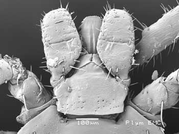
Nymph - dorsal view of the capitulum,
chelicerae at sides, behind hypostome tip |
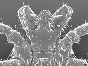
Nymph - ventral view of the hypostome,
chelicerae not visible |
 |
 |
Male - dorsal view of the capitulum, showing the
smooth surface of the hypostome, pedipalps and
rudimentary porose areas? |
Male - ventral view of capitulum, with hypostome
and chelicerae laterally (different form to female) |
 |
 |
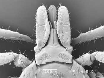
Female - dorsal view - showing capitulum
with
central hypostome, chelicerae at the sides,
largely obscured by the pedipalps.
Female has pronounced
porose areas for applying glue to eggs |
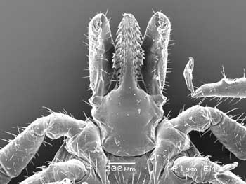
Female - ventral view of female capitulum,
note
chelicerae largely obscured at sides of hypostome |