|
|
Dartmoor Tick Watch
|
|
| Home | Collecting kit | Record sheet | Results | Disease statistics | Photos | Tick ID | Safety | News | Thanks | Contact |
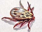
Sheep tick - Ixodes ricinus
FEMALE
Click here for: larva, nymph, male, female, mating ticks and comparing mouthparts photographs
![]()
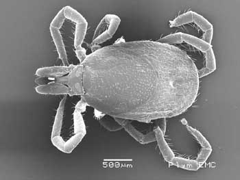
These photos were taken on the JEOL 5600LVSEM
scanning electron microscope at the University of Plymouth Electron Microscopy
Centre on 10th August 2009.
![]() |
![]() |
|
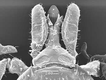
Dorsal view - adult female
|
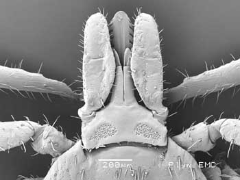
Dorsal view - showing capitulum with
central hypostome, chelicerae, pedipalps and porose areas
|
![]() |
![]() |
|
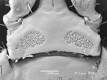
Porose areas with lateral cornuae
|
Similar dorsal view of another female
|
![]() |
![]() |
|
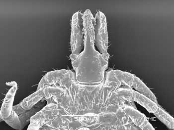
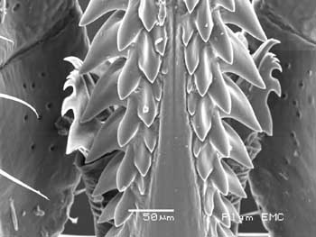
Ventral view of female capitulum
|
As at left - debris on the hypostome
|
![]() |
![]() |
|
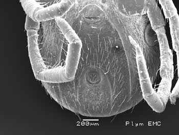
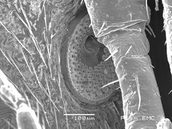
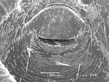
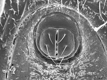
Female aperture (top) and anus (below)
|
Spiracle, partly hidden
|
![]() |
![]() |
|
Female aperture
|
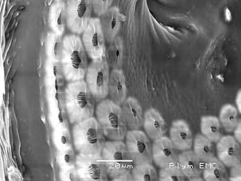
Spiracle detail
|
![]() |
![]() |
|
Anus
|
Ventral view of female capitulum, note
chelicerae largely obscured
|
![]() |
![]() |
|
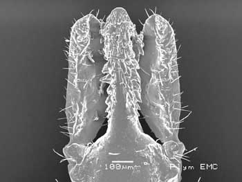
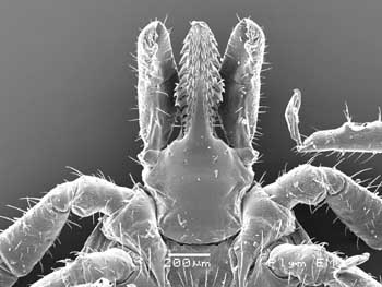
Anterior view of hypostome with chelicerae
at sides
|
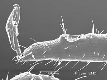
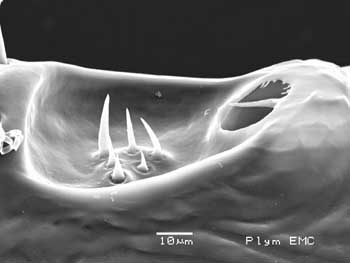
Left leg 1, with Haller's organ, note extra
pores towards the foot
|
![]() |
![]() |
|
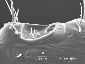
Haller's organ, with sensillae (hairs) and
sensory pit
|
Haller's organ detail
|
Acknowledgement: My thanks to the staff of the University of Plymouth Electron Microscopy Centre, Dr Roy Moate, Peter Bond and Glenn Harper, for enabling me to take these photographs.